5/4 Blog Post

On Thursday, our class toured the University of Kentucky's Electron Microscopy Center. However, due to the NCAA tournament in Lexington, Virginia, I was unable to attend. However, based on what our professors informed us about, and what I heard from my fellow classmates, three different types of microscopy were discussed: Scanning Electron Microscopy (SEM), Focused Ion Beam Microscopy, and Energy Dispersive Spectroscopy. I am disappointed that I was unable to attend the field trip, as these devices have not only enabled so many advancements in scientific fields, but will continue to do so.
I would first like to start with a discussion of what I learned from Chapter 5 in our book Size Really Does Matter by Colm Durkan. As a biology student, microscopes have played an utmost important role in my laboratory experiences. Specifically, this last semester as a microbiology student, I was using microscopes and relying on their magnification three times a week. The main difficulty or limit that light microscopes face is the wavelength of light. However, the curvature and size of the lens can control the image. Microscopes not only have to magnify, but they have to produce quality resolution so that the user can observe whatever they are looking at. Empty magnification is magnification at a low resolution. One can magnify something as many times as they would like, but if they cannot get a clear resolution, that magnification surmounts to nothing. Another issue that presents itself is the refractive qualities of light, specifically as it travels through air. The more air the light travels through, the less focused the light truly is. As very amateur microbiologists, we used oil immersion. The oil has a similar refractive index as the glass of the slide, therefore, there is less refraction and less distortion on the image. However, this still is not enough. Therefore, we turned to things other than light for microscopy.
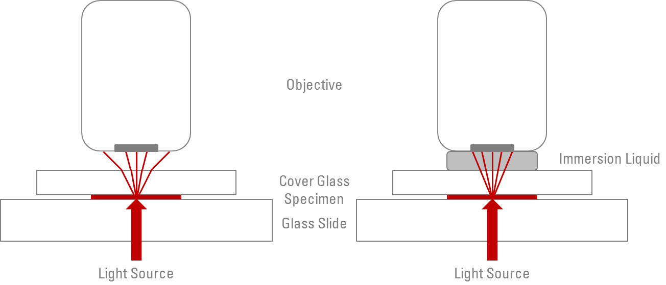
F. 1 This picture shows the increase of focused light with oil immersion! This is somethingthat I was unaware of until Microbiology, and was excited that I finally learned the physics behind why we use oil.
Scanning electron microscopes are relatively simple. A single beam of electrons is shone onto the sample. The electrons then interact with the sample, scattering the electrons, secondary electrons (which are electrons produced by ionization), etc. These signals are then collected by detectors and form an image on the computer screen. The electron beam is shone using a raster scan process —rectangular scanning process. The signal received on the detectors is dependent on the sample thickness and atomic composition. Everything interacts slightly differently with the electron beam. Scanning electron microscopes cannot produce atomic resolution, but they can get remarkably close (around 1-2 nm).
The Focused Ion Beam microscopy is done through scanning electron microscopes. The most important application of this technique is with metals and determining their inter-metallic interactions and formations. This device takes all of the designs of a scanning electron microscope but adds a second beam. The second beam then cuts into the sample, effectively destroying it. This is a large downside or limitation to FIB-SEM, the sample is destroyed after analysis. However, the huge reward is 3D analysis of the sample rather than 2D, which is all that SEM can do. Focused Ion Beam allows for precise cross sectioning, and exposure of the entire sample rather than just the surface. Therefore, I would imagine that FIB-SEM is used with unknown samples that are many in number. It would be used in a circumstance where a large quantity of information needs to be determined about the sample, and the user has access to a plethora of that sample. Yet, what happens if every sample is slightly different? If the microscopy damages the sample, for this example's sake, entirely beyond repair, how does one ensure that the next sample is identical and apply the knowledge learned from the destroyed sample A to the usable sample B? Unfortunately, I could not attend the tour or else I would have been sure to ask that question, but perhaps that question underlies most of science...

Energy Dispersive Spectroscopy is also done through scanning electron microscopes. This time, however, the secondary electrons, backscattered electrons, and X-Rays are used to quantify chemical composition. It has a detection limit at 0.1% so it cannot detect any element in trace amounts, but any surplus quantity can be detected by the EDS system. Energy Dispersive Spectroscopy relies on Moseley's Law which states that each type of atom (each element) has a distinct frequency of limit emitted when core electrons are ejected. X-Rays are used to eject these core electrons which then emits a signature frequency for the specific element it came from. One drawback to EDS is that it cannot be used to quantify low atomic number elements due to the lack of abundance of outer electrons. However, unlike FIB it does not destroy the sample.

As described above, each spectroscopy has its own specialty. I would have loved to been able to see these first hand at the UK Center, but enjoyed researching about these pieces of equipment and how they are able to be used. From a nano-science perspective, these devices are incredibly useful, allowing researchers to see samples on a 2D or 3D level, and to determine chemical composition. While I am unsure of the actual healthcare applications, I can imagine that these could be incredibly useful in identifying how to destroy unknown pathogens. If the membrane composition can be determined of a pathogen, finding a cure becomes more manageable. Overall, I have been fascinated learning about the different equipment that makes nano-science possible, and am looking forward to continuing to learn more!
Works Cited:
“Energy-Dispersive X-Ray Spectroscopy (EDS).” Chemistry LibreTexts, 22 Aug. 2022, chem.libretexts.org/Courses/Franklin_and_Marshall_College/Introduction_to_Materials_Characterization__CHM_412_Collaborative_Text/Spectroscopy/Energy-Dispersive_X-ray_Spectroscopy_(EDS).
“Focused Ion Beam Scanning Electron Microscopy (FIB-SEM).” Covalent Metrology Analytical Labs, 28 July 2022, covalentmetrology.com/techniques/focused-ion-beam-scanning-electron-microscopy-fib-sem/#:~:text=Like%20other%20high%2Dresolution%20scanning,features%20on%20a%20sample%20surface.
“Scanning Electron Microscopes.” Electron Microscopy Center, emc.engr.uky.edu/equipment/equipment-list/scanning-electron-microscopes. Accessed 8 May 2023.
Comments
Post a Comment